
- MathNotebook
- MathConcepts
- StudyMath
- Geometry
- Logic
- Bott periodicity
- CategoryTheory
- FieldWithOneElement
- MathDiscovery
- Math Connections
Epistemology
- m a t h 4 w i s d o m - g m a i l
- +370 607 27 665
- My work is in the Public Domain for all to share freely.
- 读物 书 影片 维基百科
Introduction E9F5FC
Questions FFFFC0
Software
Physics discovery, Math discovery, Neuroscience Notes
Lapkričio mėn. 29 d. Behavioural Medicine and Neuroplasticity, Kaunas, VDU, S.Daukanto g. 28. Pakabinti 8:30-9:30. Numeris: P16.
A0 (841 x 1189 mm), portretas. 16 lapų.
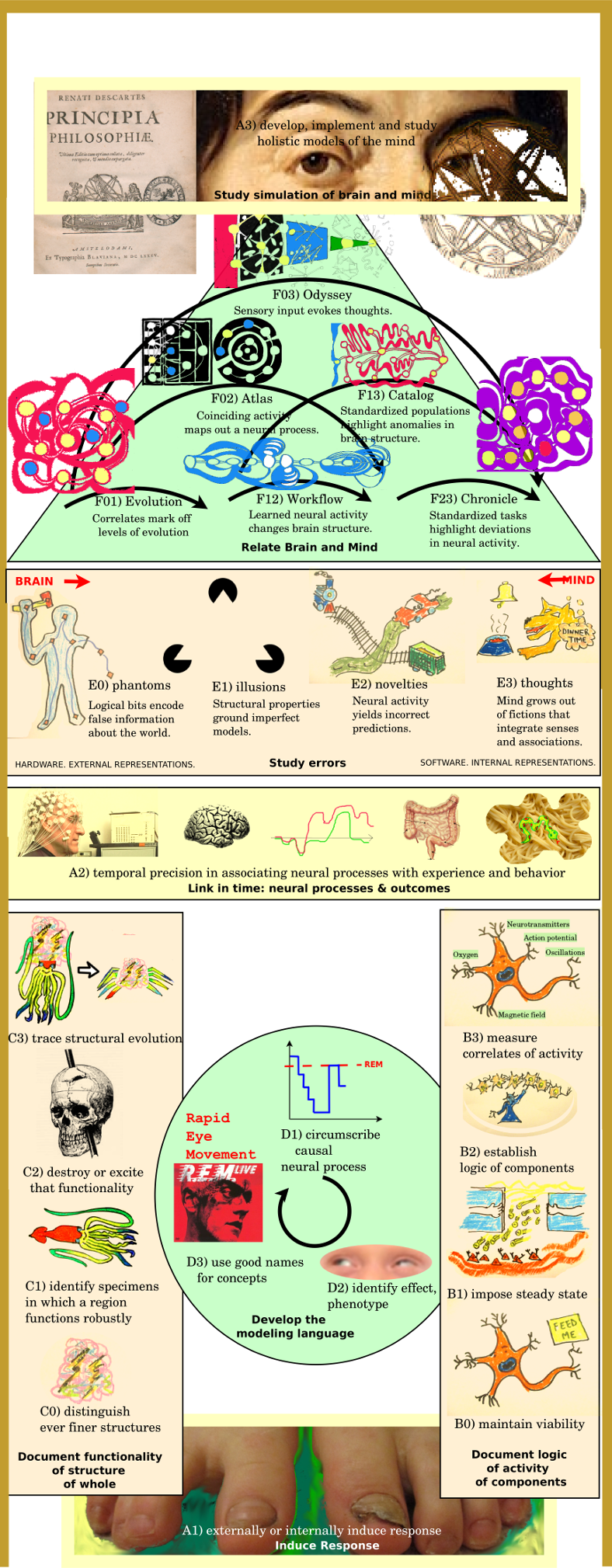
Expressing the Organizer of an Organism's Behavior
An Epistemological Overview of Neuroscience
Darbai
- Išryškinti, išmąstyti ir pavaizduoti šešis pertvarkymus.
- Palyginti keturias klaidas su tiesos apibrėžimais.
- Patikslinti visus išsiaiškinimo būdus.
- Parinkti paveikslėlių, brėžinių.
- Aprašyti tyrimą.
- Surašyti išvadų santrauką.
- What does it mean for the organism to have an internal impulse (from its mind) to which it responds?
- Where do the levels of awareness of that mind come into play? leading to its model of itself, its awareness and consciousness?
- In what sense does the brain generate a mind as its will, executor, controller, god?
- How do simulations bring out the distinction between brain and mind?
- Is there an important difference between externally and internally induced brain activity? Does one get interesting results if one trains oneself to modify one's functional neural image?
Description of Investigation
Main conclusions
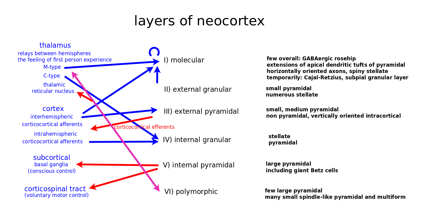
Perspective is the inversion of mind with regard to brain. Perspective of perspective is a second inversion that has feelings convey the brain to the mind. Perpsective of perspective of perspective is a third inversion that establish an independent reference point from which to choose how to adjust the mind body relationship. Hypothesis: Six layer cortex.
The binding problem: How does information come all back together.
An organism, by definition, is that which has a "purpose" - to persist - and is organized to support that purpose. It is part of an environment, an ecosystem that likewise supports that purpose. And then that purpose manifests itself through an organizer for the organism - the brain - and through "thoughts" that are "harmonies" (solutions) that form the basis for the mind, which is the dual inverse of the brain, derived from those nonphysical harmonies.
Study of Six Visualizations
Visualizations compare "modifications" to the "standardized" brain. Which is to say, there can be variations of a working model. The working model is representative of a population, and the individual represents a variation upon that model.
Visualization relates "what it is" with "how it adapts to the world". So the facts state what it is, (the hardware), and the thoughts are just adaptations (the software). The mind is an idealistic reinterpretation whereby the set of thoughts are "what it is". The software becomes the hardware.
- Evolution: Tree of evolution of populations organized with a sequence of stages with structural properties characteristic of more sophisticated models.
- Interpretation of correlates to evolution.
- Atlas: Network of predictive activity of neural processes organized into hierarchy of tasks.
- Functional Map.
- Handbook: (Underlying structural support for) Sequences of events (internal tasks) (in parallel) (facilitated) is reorganized (rewired) by network of structure (structural changes) to support confirmed predictions (about external world)
- Observed change in structure (learning behavior)
- Chronicle: Sequence of behavior reorganized by a hierarchy of tasks.
- Neural expression of standardized behavior.
- Catalog: Hierarchy of standard modules reorganized with network of links (alternate routes).
- Neural expression of a standardized human conditions.
- Odyssey: Network of signal flow organized with a sequence.
- Causal network: Follow the progress of sensory input.
Notes
Key words
Other methods
Compare liminal and subliminal responses (fear of snakes).
Unspecified task - rest state - natural activity "in the field".
Importance of twin studies
- 50% of traits attributed to genetics. But little attributed to family environment. Thus development is an important source.
Decoding
- Use artificial intelligence to recognize patterns. James V. Haxby
- Reconstruct experience from brain activity.
Have people think at rest
- Correlate with fMri
Logical fallacies
- Reverse inference. Just because the amygdala is active doesn't mean that the person is experiencing fear.
Overall Idea
Also, establishing a map between the brain and the mind, whereby the brain is a system that implements the mind.
Relationship of mind and brain
- David Rioch helped create one of the first interdisciplinary psychiatric research programs at Walter Reed, composed of two interacting groups of scientists, a behavioral group and a brain group. David Hubel has written that "In the neuropsychiatry division, David Rioch had assembled a broad and lively group of young neuroscientists ... the focus was on the entire nervous system, not on a subdivision of biological subject matter based on methods."
- The first freestanding neuroscience department (then called Psychobiology) was founded in 1964 at the University of California, Irvine by James L. McGaugh.
- Francis O. Schmitt established a neuroscience research program within the Biology Department at the Massachusetts Institute of Technology, bringing together biology, chemistry, physics, and mathematics.
- Brain research has gone through philosophical, experimental, and theoretical phases.
Externally induce response
Externally: can be by the person themselves. They are external to their own brain.
Externally Induce Response - Typically using two locations.
- Frog galvanoscope Sensitivity to external potential. Frog's leg muscle will twitch when the sciatic nerve is included in an eletric circuit (by making two electrical connections with it) and when removed from it.
- Luigi Galvani Galvani's assistant touched an exposed sciatic nerve of the frog with a metal scalpel that had picked up a charge. At that moment, they saw sparks and the dead frog's leg kicked as if in life. The observation made the Galvanis the first investigators to appreciate the relationship between electricity and animation—or life.
- Motor NCS are performed by electrical stimulation of a peripheral nerve and recording from a muscle supplied by this nerve. The time it takes for the electrical impulse to travel from the stimulation to the recording site is measured.
- Sensory NCS are performed by electrical stimulation of a peripheral nerve and recording from a purely sensory portion of the nerve, such as on a finger.
- The scientists held a sheathed coil of wire against the scalp and “zapped” it—sent an intense pulse of magnetic energy into the skull—inducing a brief electric current in the neurons underneath. The perturbation, in turn, excited and inhibited the neurons’ partner cells in connected regions, in a chain reverberating across the cortex, until the activity died out. A network of electroencephalogram (EEG) sensors, positioned outside the skull, recorded these electrical signals. As they unfolded over time, these traces, each corresponding to a specific location in the brain below the skull, yielded a movie.These unfolding records neither sketched a stereotypical pattern, nor were they completely random. Remarkably, the more predictable these waxing and waning rhythms were, the more likely the brain was unconscious. The researchers quantified this intuition by compressing the data in the movie with an algorithm commonly used to “zip” computer files. The zipping yielded an estimate of the complexity of the brain’s response. Volunteers who were awake turned out have a “perturbational complexity index” of between 0.31 and 0.70, dropping to below 0.31 when deeply asleep or anesthetized. Massimini and Tononi tested this zap-and-zip measure on 48 patients who were brain-injured but responsive and awake, finding that in every case, the method confirmed the behavioral evidence for consciousness. The team then applied zap and zip to 81 patients who were minimally conscious or in a vegetative state. For the former group, which showed some signs of nonreflexive behavior, the method correctly found 36 out of 38 patients to be conscious. It misdiagnosed two patients as unconscious. Of the 43 vegetative-state patients in which all bedside attempts to establish communication failed, 34 were labeled as unconscious, but nine were not. Their brains responded similarly to those of conscious controls—implying that they were conscious yet unable to communicate with their loved ones.
Demonstrated lack of correlation
- Refutation of Balloonist theory Volume of muscle did not increase when caused to contract (Jan Swammerdam), or when flexed (Francis Glisson).
Study ensembles
- Record and stimulate activity - for single neurons and multiple neurons and networks.
Activate region
- Stimulate physically
- Stimulate mentally
Neurostimulation The purposeful modulation of the nervous system's activity using invasive (e.g. microelectrodes) or non-invasive means (e.g. transcranial magnetic stimulation or transcranial electric stimulation, tES, such as tDCS or transcranial alternating current stimulation, tACS). Neurostimulation usually refers to the electromagnetic approaches to neuromodulation.
- Luigi Galvani discovered that the muscles of dead frog legs twitched when struck by direct current on the nervous system.
- Delgado used stimulation as an experimental manipulation to study basics of how the brain works. The primary works were on the reward center of the brain in which stimulation of those structures led to pleasure that requested more stimulation.
- The electrical stimulation of the MT area of primary visual cortex biases perception. In particular, the directionality of motion is represented in a regular way in the MT area. They presented monkeys with moving images on screen and monkey throughput was to determine what the direction is. They found that by systematically introducing some errors to the monkey's responses, by stimulating the MT area which is responsible for perceiving the motion in another direction, the monkey responded to somewhere in between the actual motion and the stimulated one. This was an elegant use of stimulation to show that MT area is essential in the actual perception of motion. * Within the memory field, stimulation is used very frequently to test the strength of the connection between one bundle of cells to another by applying a small current in one cell which results in the release of neurotransmitters and measuring the postsynaptic potential.
Three-cycle
Name concepts
- If you don't use names for concepts, then you can't develop the right theory. If you use good names, then the theory develops straightforwardly.
Parcellation of brain
- It is highly desirable to parcellate the brain into functionally distinct parcels: brain regions with distinct architectonics, connectivity, function, and/or topography (Felleman and Van Essen, 1991).[14] Accurate parcellation allows each node in the macroscale connectome to be more informative by associating it with a distinct connectivity pattern and functional profile. Parcellation of localized areas of cortex have been accomplished using diffusion tractography (Beckmann et al. 2009)[35] and functional connectivity (Nelson et al. 2010)[36] to non-invasively measure connectivity patterns and define cortical areas based on distinct connectivity patterns. Such analyses may best be done on a whole brain scale and by integrating non-invasive modalities. Accurate whole brain parcellation may lead to more accurate macroscale connectomes for the normal brain, which can then be compared to disease states.
Flexible regions that change allegiance.
- Modulate flexibility by fatigue, mood, arousal, drugs.
- Identifying network phenotype - features of high performing, well learning individuals.
- Need multivariate behavior models.
- Bridge network descriptors of brain with univariate activation descriptors of brain.
- Bridge network descriptors of brain with models of behavior.
- Generative models and theories of network dynamics.
- Annotative graph.
Document activity
Maintain viability
Cell culture is the process by which cells are grown under controlled conditions, generally outside their natural environment. After the cells of interest have been isolated from living tissue, they can subsequently be maintained under carefully controlled conditions. These conditions vary for each cell type, but generally consist of a suitable vessel with a substrate or medium that supplies the essential nutrients (amino acids, carbohydrates, vitamins, minerals), growth factors, hormones, and gases (CO2, O2), and regulates the physio-chemical environment (pH buffer, osmotic pressure, temperature).
Activity at rest.
- Maintain viability, functionality - keep alive.
- Observation of activity when subject is doing nothing
Impose steady state
Maintain constant voltage or current
- Voltage clamp The voltage clamp allows the membrane voltage to be manipulated independently of the ionic currents, allowing the current-voltage relationships of membrane channels to be studied. Cell membranes of excitable cells contain many different kinds of ion channels, some of which are voltage-gated. Voltage-gated ion channels are a class of transmembrane proteins that form ion channels that are activated by changes in the electrical membrane potential near the channel. The membrane potential alters the conformation of the channel proteins, regulating their opening and closing.
- Electrophysiology is the branch of physiology that pertains broadly to the flow of ions (ion current) in biological tissues and, in particular, to the electrical recording techniques that enable the measurement of this flow. Classical electrophysiology techniques involve placing electrodes into various preparations of biological tissue.
- If an electrode is small enough (micrometers) in diameter, then the electrophysiologist may choose to insert the tip into a single cell. Such a configuration allows direct observation and recording of the intracellular electrical activity of a single cell. However, this invasive setup reduces the life of the cell and causes a leak of substances across the cell membrane.
- Patch clamp technique is a laboratory technique in electrophysiology used to study ionic currents in individual isolated living cells, tissue sections, or patches of cell membrane. Patch clamping can be performed using the voltage clamp technique. In this case, the voltage across the cell membrane is controlled by the experimenter and the resulting currents are recorded. Alternatively, the current clamp technique can be used. In this case the current passing across the membrane is controlled by the experimenter and the resulting changes in voltage are recorded, generally in the form of action potentials.
- Galvanometer is an electromechanical instrument used for detecting and indicating an electric current. It was essential to discovering the electrical activity of the heart and brain, by its fine measurements of current.
- Electrophysiology is the branch of physiology that studies the electrical properties of biological cells and tissues. It involves measurements of voltage changes or electric current or manipulations on a wide variety of scales from single ion channel proteins to whole organs like the heart. In neuroscience, it includes measurements of the electrical activity of neurons, and, in particular, action potential activity. Recordings of large-scale electric signals from the nervous system, such as electroencephalography, may also be referred to as electrophysiological recordings
- Action potential: Experimental methods Alan Lloyd Hodgkin and Andrew Fielding Huxley focused on three goals: isolating signals from single neurons or axons, developing fast, sensitive electronics, and shrinking electrodes enough that the voltage inside a single cell could be recorded.
- The first problem was solved by studying the giant axons found in the neurons of the squid (Loligo forbesii and Doryteuthis pealeii, at the time classified as Loligo pealeii). These axons are so large in diameter (roughly 1 mm, or 100-fold larger than a typical neuron) that they can be seen with the naked eye, making them easy to extract and manipulate.
- The second problem was addressed with the crucial development of the voltage clamp, which permitted experimenters to study the ionic currents underlying an action potential in isolation, and eliminated a key source of electronic noise, the current IC associated with the capacitance C of the membrane. Since the current equals C times the rate of change of the transmembrane voltage Vm, the solution was to design a circuit that kept Vm fixed (zero rate of change) regardless of the currents flowing across the membrane.
- The third problem, that of obtaining electrodes small enough to record voltages within a single axon without perturbing it, was solved in 1949 with the invention of the glass micropipette electrode, which was quickly adopted by other researchers.
- While glass micropipette electrodes measure the sum of the currents passing through many ion channels, studying the electrical properties of a single ion channel became possible in the 1970s with the development of the patch clamp by Erwin Neher and Bert Sakmann. For this discovery, they were awarded the Nobel Prize in Physiology or Medicine in 1991. Patch-clamping verified that ionic channels have discrete states of conductance, such as open, closed and inactivated.
- Optical imaging technologies have been developed in recent years to measure action potentials, either via simultaneous multisite recordings or with ultra-spatial resolution. Using voltage-sensitive dyes, action potentials have been optically recorded from a tiny patch of cardiomyocyte membrane.
Establish functional components and their logic
Reveal units and their structure
- Golgi's method A silver staining technique that is used to visualize nervous tissue under light microscopy. It stains cells randomly, allowing their structure to be distinguished from the mass of surrounding structure. This makes it possible to classify cell types, and fiber types. This let Cajal disprove the reticular theory, despite Golgi's support for it. That theory stated that the brain is one continuous network. Cajal showed that the relationship between neurons is not continuous but continguous. Thus there are units of information processing.
Alter activity.
Research in 2004 has shown that synapses do not strengthen or weaken on a sliding scale. There are discrete states that synapses move between. These states are active, silent, recently silent, potentiated, and depressed. The states which they can move to are dependent on the state that they are in at the moment. Thus, the future state is determined by the state gained by previous activity. For instance, silent (but not recently silent) synapses can be converted to active via the insertion of AMPARs in the postsynaptic membrane. Active synapses can move to either potentiated or depressed via LTP or LTD respectively. Prolonged low-frequency stimulation (5 Hz, the method used to induce LTD) can move an active synapse to depressed and then silent. However, synapses that have just become active cannot be depressed or silenced. Thus there is state-machine-like behavior at the synapse when it comes to transitions. However, the states themselves can have varying degrees of intensity. One active-state synapse can be stronger than another active-state synapse. This is, in theory, how you can have a strong memory vs. a weak memory. The strong memories are the ones with very heavily populated active synapses, while weak memories may still be active but poorly populated with AMPARs. The same research has shown that NMDA receptors themselves, once thought to be the control mechanism behind AMPA receptor organization, can be regulated by synaptic activity. This regulation of the regulation mechanism itself adds another layer of complexity to the biology of the brain. (Metaplasticity)
Neuromodulation (therapy) The alteration of nerve activity through targeted delivery of a stimulus, such as electrical stimulation or chemical agents, to specific neurological sites in the body. It is carried out to normalize – or modulate – nervous tissue function.
- Neuromodulation The physiological process by which a given neuron uses one or more chemicals to regulate diverse populations of neurons.
Introduce hormones or neurotransmitters to cell culture (?)
Establish correlates of activity
Correlates of activity are the orders of magnitude.
Correlates can include oxygen but also neurotransmitters.
Document Structure
Distinguish ever finer regions
Structural imaging.
- CT Scans (Computed Tomography).
- Magnetic Resonance Imaging. (MRI).
- Neuroimaging or brain imaging The use of various techniques to either directly or indirectly image the structure, function, or pharmacology of the nervous system.
- Structural imaging deals with the structure of the nervous system and the diagnosis of gross (large scale) intracranial disease (such as a tumor) and injury.
- Functional imaging is used to diagnose metabolic diseases and lesions on a finer scale (such as Alzheimer's disease) and also for neurological and cognitive psychology research and building brain-computer interfaces.
- Magnetic resonance imaging Magnetic resonance imaging is a medical application of nuclear magnetic resonance (NMR). Hydrogen atoms are most often used to generate a detectable radio-frequency signal that is received by antennas close to the anatomy being examined. Hydrogen atoms are naturally abundant in people and other biological organisms, particularly in water and fat. For this reason, most MRI scans essentially map the location of water and fat in the body.
- MRI sequence A particular setting of pulse sequences and pulsed field gradients, resulting in a particular image appearance.
- MRI contrast agent improves visibility.
- Diffusion-weighted magnetic resonance imaging The use of specific MRI sequences as well as software that generates images from the resulting data that uses the diffusion of water molecules to generate contrast in MR images. Molecular diffusion in tissues is not free, but reflects interactions with many obstacles, such as macromolecules, fibers, and membranes. Water molecule diffusion patterns can therefore reveal microscopic details about tissue architecture, either normal or in a diseased state.
- Tractography. Aa 3D modeling technique used to visually represent nerve tracts using data collected by diffusion MRI. It uses special techniques of magnetic resonance imaging (MRI) and computer-based diffusion MRI. The results are presented in two- and three-dimensional images called tractograms.
- CT scan Computed tomography scan. Makes use of computer-processed combinations of many X-ray measurements taken from different angles to produce cross-sectional (tomographic) images (virtual "slices") of specific areas of a scanned object, allowing the user to see inside the object without cutting. CT produces data that can be manipulated in order to demonstrate various bodily structures based on their ability to absorb the X-ray beam.
- Connectogram graphical representations of connectomics, the field of study dedicated to mapping and interpreting all of the white matter fiber connections in the human brain. These circular graphs based on diffusion MRI data utilize graph theory to demonstrate the white matter connections and cortical characteristics for single structures, single subjects, or populations.
Finding shared features
- General features across modules: dense connectivity.
Differentiating different parts of the brain
- Dissection of human brain. Including post-mortem analysis of various diseases.
- Cytoarchitecture Viennese psychiatrist Theodor Meynert (1833–1892) in 1867 noticed regional variations in the histological structure of different parts of the gray matter in the cerebral hemispheres.
- Constantin von Economo and Georg N. Koskinas, two neurologists in Vienna, produced a landmark work in brain research by defining 107 cortical areas on the basis of cytoarchitectonic criteria.
- Nissl staining technique. Nissl staining reveals macroscopic details such as the laminar pattern of the cerebral cortex or the interlocking nuclear patterns of the diencephalon and brainstem.
- Nissl staining reveals microscopic details such as the distinctions between individual neurons and glia in any subregion of the central nervous system.
Identify specimens (or populations) in which a region functions robustly or poorly
Dissection of human brain
- Regularities and irregularities. What is normal?
Healthy people as a baseline.
- Savant syndrome, Genius, Intellectual giftedness Study people with exceptional abilities.
Destroy that functionality
Remove as little as possible to destroy function (lowest upper bound), or as much as possible and yet maintain function (greatest lower bound).
- Manipulate the Brain. Pharmacological lesioning. Use drugs to stop the brain from working in certain ways. Organic amnesia. Amnesia while conscious. Conscious vs. unconscious components of memory
- Transcranial magnetic stimulation. Shut off language production center - Broca's area.
- Physical lesioning in animals
- Specific neurosurgeries in humans - small removals
Effect of Absence
- The Roman physician Galen, a follower of Hippocrates and physician to Roman gladiators, observed that his patients lost their mental faculties when they had sustained damage to their brains.
- Roger W. Sperry shows the potential independence of the two sides of the human brain using split-brain patients (1962–1965).
- Jean Pierre Flourens pioneered the experimental method of carrying out localized lesions of the brain in living animals describing their effects on motricity, sensibility and behavior.
- Work with brain-damaged patients by Paul Broca suggested that certain regions of the brain were responsible for certain functions. At the time, Broca's findings were seen as a confirmation of Franz Joseph Gall's theory that language was localized and that certain psychological functions were localized in specific areas of the cerebral cortex.
- The localization of function hypothesis was supported by observations of epileptic patients conducted by John Hughlings Jackson, who correctly inferred the organization of the motor cortex by watching the progression of seizures through the body. Carl Wernicke further developed the theory of the specialization of specific brain structures in language comprehension and production.
Inspection of tissue
- Brain biopsy The removal of a small piece of brain tissue for the diagnosis of abnormalities of the brain. It is used to diagnose tumors, infection, inflammation, and other brain disorders. Various brain abnormalities can be diagnosed by microscopic analysis of the tissue sample. The pathologist (a physician trained in how disease affects the body's tissues) looks for abnormal growth, changes in cell membranes, and/or abnormal collections of cells. In Alzheimer's disease, the cortex of the brain contains abnormal collections of plaques. If infection is suspected, the infectious organism can be cultured from the tissue and identified.
Effect of Deletion
- Ablative brain surgery By ablating specific brain regions and observing differences in animals subjected to behavioral tests, the functions of all the removed areas may be inferred.
- The effects of brain lesions (caused by accidents or diseases) on behavior can be observed to draw conclusions on the functions of different parts of the brain.
- Pallidotomy Destruction of a small area of brain cells by heating a tiny electrical probe.
- César Julien Jean Legallois He began a long series of physiological experiments to study the basic physical conditions necessary for the maintenance of life functions throughout the organism. Legallois conducted a series of animal experiments to clarify the mechanism of respiration. By decapitation of vertebrates or other targeted destruction of neural connections in the brain and spinal cord, he came to the conclusion that respiration is controlled by a respiratory center located in the medulla oblongata. His discovery was that a lesion, on a small circumscribed area in the medulla, inhibits breathing (1811). This was the first attempt to localize respiratory regulation and was later completed by the work of Marie-Jean-Pierre Flourens (1794-1867).
- César Julien Jean Legallois The demonstration of metameric organization of the spinal cord, from which each segment as a neural center of a particular region (e.g. As dermatome, Myotom) serves to coordinate about their sensory and motor activity.
Destructive interference
- Deep brain stimulation Placing a neurostimulator (a "brain pacemaker") which sends electrical impulses to alter brain activity, for example, to block abnormal behavior.
Transcranial magnetic stimulation A noninvasive form of brain stimulation in which a changing magnetic field is used to cause electric current at a specific area of the brain through electromagnetic induction. An electric pulse generator, or stimulator, is connected to a magnetic coil, which in turn is connected to the scalp. The stimulator generates a changing electric current within the coil which induces a magnetic field; this field then causes a second inductance of inverted electric charge within the brain itself.
- Single or paired pulse TMS causes neurons in the neocortex under the site of stimulation to depolarize and discharge an action potential.
- If used in the primary motor cortex, it produces muscle activity referred to as a motor evoked potential (MEP) which can be recorded on electromyography.
- If used on the occipital cortex, 'phosphenes' (flashes of light) might be perceived by the subject.
- In most other areas of the cortex, there is no conscious effect, but behaviour may be altered (e.g., slower reaction time on a cognitive task), or changes in brain activity may be detected using diagnostic equipment.
- Repetitive TMS produces longer-lasting effects which persist past the period of stimulation. rTMS can increase or decrease the excitability of the corticospinal tract depending on the intensity of stimulation, coil orientation, and frequency. Low frequency rTMS with a stimulus frequency less than 1 Hz is believed to inhibit cortical firing while a stimulus frequency greater than 1 Hz, or high frequency, is believed to provoke it.[23] Though its mechanism is not clear, it has been suggested as being due to a change in synaptic efficacy related to long-term potentiation (LTP) and long-term depression (LTD).
- Electromyography An electrodiagnostic medicine technique for evaluating and recording the electrical activity produced by skeletal muscles.
Replace with stimulator, or implant stimulator
- "Your face just melted."
In Vivo electrical stimulation
- In humans during neurosurgery - stimulate parts of the brain with electricity - to map out brain to protect it or investigate it
- In animals
Trace structural evolution
Dissection of animal brain - Comparisons
- David Rioch demonstrated in detail for the first time how the forebrains of dogs and cats are more complex than those of rodents.
Observation of the development of the nervous system in animals and humans.
- Observe evolution of brain during life cycle of individual.
- Evolution of the nervous system across species. Comparison of different species.
Temporal state
Observation of brain activity
- Electroencephalography records electrical activity of the brain, typically through electrodes placed along the scalp.
- Measuring electric activity: Electroencephalogram (EEG). State the brain is in.
- Good temporal resolution, lousy spatial resolution.
- Electroencephalographic tasks.
- Event-related potentials (ERP) are
- Time-locked recording of electrical activity.
- Spectral content of EEG.
Observed variation of signal strength
- Richard Caton used a galvanometer with one electrode attached to the cerebral cortex and another to the surface of the skull. He noticed the variation of the signal strength with regard to different behaviors, such as sleep or the onset of death.
Time taken to complete task. Accuracy.
- Compare nonwords, words, pseudowords.
Performance of standardized behavior
Mental chronometry (reaction time studies) - use of response time in perceptual-motor tasks to infer content, duration and temporal sequencing of mental/cognitive operations.
- Subtraction - reaction time for additional decision
- Simple reaction time < Recognition reaction time < Choice reaction time
- Sternberg: Recognition time ~ N for number of items
- Hick's law: Choice reaction time ~ log N
Electrodiagnosis (EDX) is a method of medical diagnosis that obtains information about diseases by passively recording the electrical activity of body parts (that is, their natural electrophysiology) or by measuring their response to external electrical stimuli (evoked potentials).[1] The most widely used methods of recording spontaneous electrical activity are various forms of electrodiagnostic testing (electrography) such as electrocardiography (ECG), electroencephalography (EEG), and electromyography (EMG).
Levels of error
What exists and does not exist. Correspondence theory of the truth. And other views of truth.
Four levels of truth given by different kinds of error
- phantoms (false information) - identified by Correspondence theory
- illusions (false models) - identified by Coherence theory
- novelties (false predictions) - identified by Construction theory
- thoughts (fictions) - identified by Consensus theory
Brain maps
- Correlation of movement with retina map - both from studies of animals where you record neurons directly - and with fMri.
Novelties: Learning behavior: Signal that modulates capacity for behavior (and thus changes behavior)
Training: Developing a conditioned stimulus
- Classical conditioning: Pavlov's research Pavlov presented a stimulus (e.g. the sound of a metronome) and then gave the dog food; after a few repetitions, the dogs started to salivate in response to the stimulus.
- Classical conditioning. Neural basis of learning and memory. classical conditioning continues to be to study the neural structures and functions that underlie learning and memory. Forms of classical conditioning that are used for this purpose include, among others, fear conditioning, eyeblink conditioning, and the foot contraction conditioning of Hermissenda crassicornis, a sea-slug. Both fear and eyeblink conditioning involve a neutral stimulus, frequently a tone, becoming paired with an unconditioned stimulus. In the case of eyeblink conditioning, the US is an air-puff, while in fear conditioning the US is threatening or aversive such as a foot shock.
Need to test capacity for behavior with one kind of signal. Then use another kind of signal to modify that capacity.
Synaptic plasticity is the ability of synapses to strengthen or weaken over time, in response to increases or decreases in their activity.
- In 1966, Terje Lømo and Tim Bliss first described the now widely studied phenomenon of long-term potentiation (LTP) in a publication in the Journal of Physiology. The experiment described was conducted on the synapse between the perforant path and dentate gyrus in the hippocampi of anaesthetised rabbits. They were able to show a burst of tetanic (100 Hz) stimulus on perforant path fibres led to a dramatic and long-lasting augmentation in the post-synaptic response of cells onto which these fibres synapse in the dentate gyrus. In the same year, the pair published very similar data recorded from awake rabbits. This discovery was of particular interest due to the proposed role of the hippocampus in certain forms of memory.
- Long-term potentiation is a persistent strengthening of synapses based on recent patterns of activity. These are patterns of synaptic activity that produce a long-lasting increase in signal transmission between two neurons. The opposite of LTP is long-term depression, which produces a long-lasting decrease in synaptic strength.
- Lømo's experiments focused on connections, or synapses, from the perforant pathway to the dentate gyrus. These experiments were carried out by stimulating presynaptic fibers of the perforant pathway and recording responses from a collection of postsynaptic cells of the dentate gyrus. As expected, a single pulse of electrical stimulation to fibers of the perforant pathway caused excitatory postsynaptic potentials (EPSPs) in cells of the dentate gyrus. What Lømo unexpectedly observed was that the postsynaptic cells' response to these single-pulse stimuli could be enhanced for a long period of time if he first delivered a high-frequency train of stimuli to the presynaptic fibers. When such a train of stimuli was applied, subsequent single-pulse stimuli elicited stronger, prolonged EPSPs in the postsynaptic cell population. This phenomenon, whereby a high-frequency stimulus could produce a long-lived enhancement in the postsynaptic cells' response to subsequent single-pulse stimuli, was initially called "long-lasting potentiation". (Long-term potentiation)
Avatar of thought
- Neurons that correspond to thoughts.
- Norman Anderson's theory - number that exists in the mind.
Visualizations
Interpretation of correlates to evolution
- Glia The ratio of glia to neurons increases through evolution, and so does the size of the glia.
Functional map
Network of neural connections, hierarchy of functionality.
Note connections between areas
- Connectome
- Human Connectome Project
- Connectome A comprehensive map of neural connections in the brain, which may be thought of as its "wiring diagram".
- Research has successfully constructed the full connectome of one animal: the roundworm Caenorhabditis elegans, which has 302 neurons. Research has explored the neural and molecular mechanisms that control several behaviors of C. elegans, including chemotaxis, thermotaxis, mechanotransduction, learning, memory, and mating behaviour.
- They proposed the development of nanoparticles that could be used as voltage sensors that would detect individual action potentials, as well as nanoprobes that could serve as electrophysiological multielectrode arrays. In particular, they called for the use of wireless, noninvasive methods of neuronal activity detection, either utilizing microelectronic very-large-scale integration, or based on synthetic biology rather than microelectronics. In one such proposed method, enzymatically produced DNA would serve as a "ticker tape record" of neuronal activity,[1][19] based on calcium ion-induced errors in coding by DNA polymerase.[20] Data would be analyzed and modeled by large scale computation.[1] A related technique proposed the use of high-throughput DNA sequencing for rapidly mapping neural connectivity.
Functional connectome
- Alivisatos et al. outlined a variety of specific experimental techniques that might be used to achieve what they termed a "functional connectome"
Related regions
- Danielle Bassett: Two regions are connected in time if they show similar patterns of activity.
Functional neuroimaging
Observation of structural activity
Neuroimaging tasks.
- Signal averaging reduces effects of extraneous activity, noise.
- Subtraction of tasks - the difference between a simpler task and a harder task is associated to a brain function. Technique in fmri.
- Positon emission tomography (PET) PET scan: tracers for blood, glucose, neurotransmitters. Blocked research designs.
- Functional MRI
- Neuroimaging
- Functional magnetic resonance imaging Measures brain activity by detecting changes associated with blood flow. Cerebral blood flow and neuronal activation are coupled.
- The primary form of fMRI uses the blood-oxygen-level dependent (BOLD) contrast to image the change in blood flow (hemodynamic response) related to energy use by brain cells. It is frequently corrupted by noise from various sources; hence, statistical procedures are used to extract the underlying signal. The resulting brain activation can be graphically represented by color-coding the strength of activation across the brain or the specific region studied. The technique can localize activity to within millimeters but, using standard techniques, no better than within a window of a few seconds.
- Arterial spin labelling
- Diffusion MRI
- Resting state fMRI, or taskless fMRI, which shows the subjects' baseline BOLD variance, revealing the existence and properties of the default mode network.
- Positron Emission Tomography PET neuroimaging is based on an assumption that areas of high radioactivity are associated with brain activity. What is actually measured indirectly is the flow of blood to different parts of the brain, which is, in general, believed to be correlated, and has been measured using the tracer oxygen-15. PET tracers emit positrons that annihilate with electrons up to a few millimeters away, causing two gamma photons to be emitted in opposite directions. PET scanner detects these emissions "coincident" in time, which provides more radiation event localization information and, thus, higher spatial resolution images than SPECT.`
- PET can be used to detect and locate chemical markers.
- Single-photon emission computed tomography (SPECT) A 2-D view of 3-D distribution of a radionuclide. Monitors level of biological activity at each place. 1 cm resolution. The nuclear isomer is taken up by brain tissue in a manner proportional to brain blood flow, which is tightly coupled to local brain metabolism and energy use.
- SPECT used to compare capacity for brain flow: Patchy loss of cortical metabolism seen in multiple strokes differs clearly from the more even or "smooth" loss of non-occipital cortical brain function typical of Alzheimer's disease.
- Fludeoxyglucose (FDG) PET scanning of the brain works to assess regional brain glucose metabolism, to provide very similar information about local brain damage from many processes.
Magnetoencephalography is a functional neuroimaging technique for mapping brain activity by recording magnetic fields produced by electrical currents occurring naturally in the brain, using very sensitive magnetometers.
- Good temporal and spatial resolution
- To generate a signal that is detectable, approximately 50,000 active neurons are needed.[9] Since current dipoles must have similar orientations to generate magnetic fields that reinforce each other, it is often the layer of pyramidal cells, which are situated perpendicular to the cortical surface, that gives rise to measurable magnetic fields. Bundles of these neurons that are orientated tangentially to the scalp surface project measurable portions of their magnetic fields outside of the head, and these bundles are typically located in the sulci.
Molecular neuroscience Various techniques for studying Neurotransmission.
- It is possible to anatomically locate neurotransmitters by labeling techniques. It is possible to chemically identify certain neurotransmitters such as catecholamines by fixing neural tissue sections with formaldehyde. This can give rise to formaldehyde-induced fluorescence when exposed to ultraviolet light. Dopamine, a catecholamine, was identified in the nematode C. elegans by using this technique.[3]
- Immunocytochemistry, which involves raising antibodies against targeted chemical or biological entities, includes a few other techniques of interest. A targeted neurotransmitter could be specifically tagged by primary and secondary antibodies with radioactive labeling in order to identify the neurotransmitter by autoradiography. The presence of neurotransmitters (though not necessarily the location) can be observed in enzyme-linked immunocytochemistry or enzyme--linked immunosorbent assays (ELISA) in which substrate-binding in the enzymatic assays can induce precipitates, fluorophores, or chemiluminescence.
- In the event that neurotransmitters cannot be histochemically identified, an alternative method is to locate them by their neural uptake mechanisms.[1]
Neural expression of a standardized human condition
Brain structure vs. Human condition
- Socioeconomic status, drug users vs. brain structure
- Neuropsychology - study disordered population - diseases (schizophrenia) and injury (stroke, aneurism, trauma).
Observed change in structure (learning behavior)
- Eric Kandel
Neuroplasticity is the ability of the brain to change continuously throughout an individual's life, e.g., brain activity associated with a given function can be transferred to a different location, the proportion of grey matter can change, and synapses may strengthen or weaken over time. The aim of neuroplasticity is to optimize the neural networks during phylogenesis, ontogeny, and physiological learning, as well as after a brain injury.
Metaplasticity refers to the plasticity of synaptic plasticity. Tthe level of synaptic inhibition, the activity of modulatory afferents such as catecholamines, and the pool of hormones affecting the synapses under study. The prior history of synaptic activity is an additional variable that influences the synaptic state, and thereby the degree, of LTP or LTD produced by a given experimental protocol. In a sense, then, synaptic plasticity is governed by an activity-dependent plasticity of the synaptic state; such plasticity of synaptic plasticity has been termed metaplasticity.
Neural expression of standardized behavior
Variation from standardized behavior related to brain structure
- Neuropsychological tests Specifically designed tasks used to measure a psychological function known to be linked to a particular brain structure or pathway. Usually involve the systematic administration of clearly defined procedures in a formal environment, free from distractions, thus offering an estimate of a person's peak level of cognitive performance. Clinical neuropsychology, Neuropsychological assessment.
Causal network: Follow the progress of sensory input
Primary receiving areas for the senses - go deeper and deeper into the senses - into levels of abstraction.
- Compare input through different senses: hearing, sight.
Optogenetics a biological technique that involves the use of light to control cells in living tissue, typically neurons, that have been genetically modified to express light-sensitive ion channels. It is a neuromodulation method that uses a combination of techniques from optics and genetics to control and monitor the activities of individual neurons in living tissue—even within freely-moving animals—and to precisely measure these manipulation effects in real-time.
- Optogenetic stimulation - directly control neurons with light.
- Diseroff at UC Davis: Genetically modify animals to put in channel redoxants, ion channels responsive to specifi frequencies of life, to label neurons, put in a fiber optic to turn the light on and off to control the flow of ions in neurons to make them fire or stop firing or fire at a certain frequency. Record and/or stimulate actual neurons. Can train an animal to do a task using light.
Note direction of signal
- Observation that more signal is going from the conceptual parts of the brain out to the sensory parts rather than the other way around.
- Jeff Hawkins emphasizes the discovery that there is ten times as much neural activity leading from the cognitive centers to the sensory centers than the other way around. This goes counter to the common understanding of the brain as a synthesizer of sensory input. Indeed, it suggest the opposite: That our mind is continuously projecting and checking absolutely everything it believes about the current reality. We are living as if in our mind's hologram.
Stimulus and response
- The idea that nerve stimulation led to movement had important implications by putting forward the idea that behaviour is based on stimuli. (Frog's leg - sciatic nerve).
- Sensory neuroscience: Single neuron experiments A typical single neuron experiment will consist of isolating a neuron (that is, navigating the neuron until the experimentor finds a neuron which spikes in response to the type of stimulus to be presented, and (optionally) determining that all of the spikes observed indeed come from a single neuron), then presenting a stimulus protocol. Because neural responses are inherently variable (that is, their spiking pattern may depend on more than just the stimulus which is presented, although not all of this variability may be true noise, since factors other than the presented stimulus may affect the sensory neuron under study), often the same stimulus protocol is repeated many times to get a feel for the variability a neuron may have. One common analysis technique is to study the neuron's average time-varying firing rate, called its post stimulus time histogram or PSTH.
- Sensory neuroscience: Receptive field estimation One major goal of sensory neuroscience is to try to estimate the neuron's receptive field; that is, to try to determine which stimuli cause the neuron to fire in what ways. One common way to find the receptive field is to use linear regression to find which stimulus characteristics typically caused neurons to become excited or depressed. Since the receptive field of a sensory neuron can vary in time (i.e. latency between the stimulus and the effect it has on the neuron) and in some spatial dimension (literally space for vision and somatosensory cells, but other "spatial" dimensions such as the frequency of a sound for auditory neurons), the term spatio temporal receptive field or STRF is often used to describe these receptive fields.
Test whether stimuli elicit responses.
- Ivan Pavlov. Reflex system research Pavlov systematically presented and withdrew stimuli to determine the antecedents that were eliciting responses, which is similar to the ways in which educational professionals conduct functional behavior assessments
- Helmholtz Time the speed at which a signal is carried along a nerve fibre.
Change components
Transfection is the process of deliberately introducing naked or purified nucleic acids into eukaryotic cells.
Infection is the invasion of an organism's body tissues by disease-causing agents, their multiplication, and the reaction of host tissues to the infectious agents and the toxins they produce.
Build simulation: Development of models
Importance of model of cognition.
- Here it is relevant to have a modular theory of cognition.
Systems analysis. Like analyzing a system of black boxes.
Structural reasoning
- Unity among duality. Pineal gland In the Passions, Descartes split man up into a body and a soul and emphasized that the soul is joined to the whole body by "a certain very small gland situated in the middle of the brain's substance and suspended above the passage through which the spirits in the brain's anterior cavities communicate with those in its posterior cavities". Descartes attached significance to the gland because he believed it to be the only section of the brain to exist as a single part rather than one-half of a pair.
Brain simulation
- Biological neuron model (Spiking neuron model) A mathematical description of the properties of certain cells in the nervous system that generate sharp electrical potentials across their cell membrane, roughly one millisecond in duration.
- Artificial neuron A mathematical function conceived as a model of biological neurons, a neural network. Artificial neurons are elementary units in an artificial neural network.
- Compartmental modelling of dendrites Helpful for understanding the behavior of neurons that are too small to record with electrodes, as is the case for Drosophila melanogaster. Neurons integrate input within dendrites, and this complexity is lost in models that assume neurons to be a fundamental unit. Dendritic branches can be modeled as spatial compartments, whose activity is related due to passive membrane properties, but may also be different depending on input from synapses.
Computational neurogenetic modeling
Other concepts
Branches of neuroscience
- Affective neuroscience Affective neuroscience is the study of the neural mechanisms involved in emotion, typically through experimentation on animal models.
- Behavioral neuroscience Behavioral neuroscience (also known as biological psychology, physiological psychology, biopsychology, or psychobiology) is the application of the principles of biology to the study of genetic, physiological, and developmental mechanisms of behavior in humans and non-human animals.
- Cellular neuroscience Cellular neuroscience is the study of neurons at a cellular level including morphology and physiological properties.
- Clinical neuroscience Clinical neuroscience The scientific study of the biological mechanisms that underlie the disorders and diseases of the nervous system.
- Cognitive neuroscience Cognitive neuroscience is the study of the biological mechanisms underlying cognition.
- Computational neuroscience Computational neuroscience is the theoretical study of the nervous system.
- Cultural neuroscience Cultural neuroscience is the study of how cultural values, practices and beliefs shape and are shaped by the mind, brain and genes across multiple timescales.
- Developmental neuroscience Developmental neuroscience studies the processes that generate, shape, and reshape the nervous system and seeks to describe the cellular basis of neural development to address underlying mechanisms.
- Evolutionary neuroscience Evolutionary neuroscience studies the evolution of nervous systems.
- Molecular neuroscience Molecular neuroscience studies the nervous system with molecular biology, molecular genetics, protein chemistry, and related methodologies.
- Neural engineering Neural engineering uses engineering techniques to interact with, understand, repair, replace, or enhance neural systems.
- Neuroanatomy Neuroanatomy the study of the anatomy of nervous systems.
- Neurochemistry Neurochemistry is the study of how neurochemicals interact and influence the function of neurons.
- Neuroethology Neuroethology Neuroethology is the study of the neural basis of non-human animals behavior.
- Neurogastronomy Neurogastronomy is the study of flavor and how it affects sensation, cognition, and memory.
- Neurogenetics Neurogenetics is the study of the genetical basis of the development and function of the nervous system.
- Neuroimaging Neuroimaging includes the use of various techniques to either directly or indirectly image the structure and function of the brain.
- Neuroimmunology Neuroimmunology is concerned with the interactions between the nervous and the immune system.
- Neuroinformatics Neuroinformatics is a discipline within bioinformatics that conducts the organization of neuroscience data and application of computational models and analytical tools.
- Neurolinguistics Neurolinguistics is the study of the neural mechanisms in the human brain that control the comprehension, production, and acquisition of language.
- Neurophysics Neurophysics deals with the development of physical experimental tools to gain information about the brain.
- Neurophysiology Neurophysiology is the study of the functioning of the nervous system, generally using physiological techniques that include measurement and stimulation with electrodes or optically with ion- or voltage-sensitive dyes or light-sensitive channels.
- Neuropsychology Neuropsychology is a discipline that resides under the umbrellas of both psychology and neuroscience, and is involved in activities in the arenas of both basic science and applied science. In psychology, it is most closely associated with biopsychology, clinical psychology, cognitive psychology, and developmental psychology. In neuroscience, it is most closely associated with the cognitive, behavioral, social, and affective neuroscience areas. In the applied and medical domain, it is related to neurology and psychiatry.
- Paleoneurobiology Paleoneurobiology is a field which combines techniques used in paleontology and archeology to study brain evolution, especially that of the human brain.
- Social neuroscience Social neuroscience is an interdisciplinary field devoted to understanding how biological systems implement social processes and behavior, and to using biological concepts and methods to inform and refine theories of social processes and behavior.
- Systems neuroscience Systems neuroscience is the study of the function of neural circuits and systems.
- molecular biology
- electrophysiology
- computational neuroscience
- Sensory neuroscience
- Stimulus
- Neural coding
Readings
General sources